Pyramidalis muscle
| Pyramidalis muscle | |
|---|---|
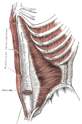 Muscles at the front of the abdomen, showing the pyramidalis at the bottom centre. | |
| Details | |
| Origin | Pubic symphysis and pubic crest |
| Insertion | Linea alba |
| Artery | Inferior and superior epigastric arteries |
| Nerve | Subcostal nerve (T12) |
| Actions | Tensing the linea alba |
| Identifiers | |
| Latin | musculus pyramidalis |
| TA98 | A04.5.01.007 |
| TA2 | 2363 |
| FMA | 15568 |
| Anatomical terms of muscle | |
The pyramidalis muscle is a small triangular muscle, anterior to the rectus abdominis muscle, and contained in the rectus sheath.
Structure
The pyramidalis muscle is part of the anterior abdominal wall.[1] Inferiorly, the pyramidalis muscle attaches to the pelvis in two places: the pubic symphysis and pubic crest, arising by tendinous fibers from the anterior part of the pubis and the anterior pubic ligament.
Superiorly, the fleshy portion of the pyramidalis muscle passes upward, diminishing in size as it ascends, and ends by a pointed extremity which is inserted into the linea alba, midway between the umbilicus and pubis.
Nerve supply
The pyramidalis muscle is innervated by the ventral portion of T12.
Blood supply
The pyramidalis is most commonly supplied by a separate branch of the inferior epigastric artery, but it may also be supplied by the superior epigastric artery in few cases. It is drained by the respective named veins.[2]
Variation
The pyramidalis muscle is present in 80% of human population.[3] It may be absent on one or both sides; the lower end of the rectus then becomes proportionately increased in size.
Occasionally, it is doubled on one side, and the muscles of the two sides are sometimes of unequal size. It may also extend higher than the usual level.
Function
The pyramidalis muscle tenses the linea alba when contracting.
Clinical significance
While making the longitudinal incision for a classical caesarean section, the pyramidalis muscle is used to determine midline and location of the linea alba.
Additional images
-
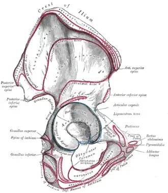 Right hip bone viewed from outside, showing a small line where the pyramidalis attaches
References
- ^ Shapiro, L. E.; Kim, J. H.; Lee, S. J.; Yoo, J. J.; Atala, A.; Ko, I. K. (2016-01-01), Lee, Sang Jin; Yoo, James J.; Atala, Anthony (eds.), "Chapter 16 - In Situ Volumetric Muscle Repair", In Situ Tissue Regeneration, Boston: Academic Press, pp. 295–312, ISBN 978-0-12-802225-2, retrieved 2021-01-23
- ^ Short, Craig L.; Crotti, Tania N.; Algate, Kent; Gladman, Marc A.; Barras, Christen D. (November 2024). "Morphology and arterial supply of the pyramidalis muscle in an Australian female population using computed tomography angiography". Surgical and Radiologic Anatomy: SRA. 46 (11): 1865–1873. doi:10.1007/s00276-024-03471-1. ISSN 1279-8517. PMC 11458779. PMID 39251450.
- ^ "7 Vestigial Features of the Human Body". Encyclopedia Britannica. Retrieved 2017-11-09.
External links
- Anatomy photo:35:11-0100 at the SUNY Downstate Medical Center - "Anterior Abdominal Wall: The Pyramidalis Muscle"
- Anatomy image:7283 at the SUNY Downstate Medical Center
- Cross section image: pelvis/pelvis-female-17—Plastination Laboratory at the Medical University of Vienna