Dog anatomy
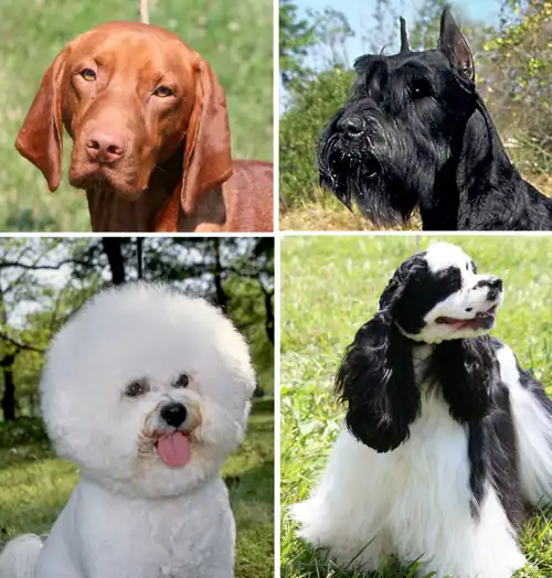
Dog anatomy comprises the anatomical study of the visible parts of the body of a domestic dog. Details of structures vary tremendously from breed to breed, more than in any other animal species, wild or domesticated,[1] as dogs are highly variable in height and weight. The smallest known adult dog was a Yorkshire Terrier that stood only 6.3 cm (2.5 in) at the shoulder, 9.5 cm (3.7 in) in length along the head and body, and weighed only 113 grams (4.0 oz). The heaviest dog was an English Mastiff named Zorba, which weighed 314 pounds (142 kg).[2] The tallest known adult dog is a Great Dane that stands 106.7 cm (42.0 in) at the shoulder.[3]
Anatomy
Muscles
The following table lists the limb muscles of the dog.[4]
| Muscle | Location | Origin | Insertion | Nerve | Action |
|---|---|---|---|---|---|
| Descending superficial pectoral | chest and shoulders | first sternebra | greater tubercle of the humerus | cranial pectoral nerves | adducts the forelimb |
| Transverse superficial pectoral | chest and shoulders | second and third sternebrae | greater tubercle of the humerus | cranial pectoral nerves | adducts the forelimb |
| Deep pectoral | chest and shoulder | ventral sternum | lesser tubercle of the humerus | caudal pectoral nerves | extends the shoulder joint |
| Sternocephalicus | chest and neck | sternum | temporal bone | accessory nerve | turns the head and neck |
| Sternohyoideus | anterior chest | Sternum | Basihyoid bone | ventral branches of the cervical spinal nerves | moves the tongue caudally |
| Sternothyoideus | neck | first costal cartilage | thyroid cartilage | ventral branches of the cervical spinal nerves | moves the tongue caudally |
| Omotransversarius | anterior back and neck | spine of the scapula | wing of the atlas | accessory nerve | advances the forelimb and flexes the neck |
| Trapezius | anterior back midline | supraspinous ligament | spine of the scapula | accessory nerve | elevates and abducts the forelimb |
| Rhomboideus | back of the neck to the shoulders | nuchal crest of the occipital bone | scapula | ventral branches of the spinal nerves | elevates the forelimb |
| Latissimus dorsi | back and shoulder | thoracolumbar fascia | teres major tuberosity of the humerus | thoracodorsal nerve | flexes the shoulder joint |
| Serratus ventralis | back of the neck to shoulders | transverse processes of the C3 to C7 vertebrae | scapula | ventral branches of the cervical spinal nerves | support the trunk and depress the scapula |
| Deltoideus | shoulder | acromial process of the scapula | deltoid tuberosity | axillary nerve | flexes the shoulder |
| Infraspinatus | shoulder | infraspinatus fossa | greater tubercle of the humerus | suprascapular nerve | extends and flexes the shoulder joint |
| Teres minor | shoulder | infra glenoid tubercle of the scapula | teres minor tuberosity of the humerus | axillary nerve | flexes the shoulder and rotates the forelimb laterally |
| Supraspinatus | shoulder | supraspinous fossa of the scapula | greater tubercle of the humerus | suprascapular nerve | extends and stabilizes the shoulder joint |
| Subscapularis | shoulder | subscapular fossa | greater tubercle of the humerus | subscapular nerve | rotates the forelimb medially |
| Teres major | shoulder | scapula | teres major tuberosity of the humerus | axillary nerve | rotates the forelimb medially |
| Coracobrachialis | shoulder | coracoid process of the scapula | crest of the lesser tubercle of the humerus | musculocutaneous nerve | adducts, extends, and stabilizes the shoulder |
| Tensor fasciae antebrachium | upper forelimb | fascia covering the latissimus dorsi | olecranon | radial nerve | extends the elbow |
| Triceps brachii | upper forelimb | caudal border of the scapula | olecranon tuber | radial nerve | extends the elbow and flexes the shoulder |
| Anconeus | lower forelimb | humerus | proximal end of the ulna | radial nerve | extends the elbow |
| Biceps brachii | upper forelimb | supraglenoid tubercle | ulnar and radial tuberosities | musculocutaneous nerve | flexes the elbow and extends the shoulder |
| Brachialis | upper forelimb | lateral surface of the humerus | ulnar and radial tuberosities | musculocutaneous nerve | flexes the elbow |
| Extensor carpi radialis | lower forelimb | supracondylar crest | metacarpals | radial nerve | extends the carpus |
| Common digital extensor | lower forelimb, carpus | lateral epicondyle of the humerus | distal phalanges | radial nerve | extends the carpus and digits 3, 4, and 5 |
| Extensor carpi ulnar | lower forelimb | lateral epicondyle of the humerus | metacarpal 5 and the accessory carpal bone | radial nerve | abducts and extends the carpal joint |
| supinator | forelimb | lateral epicondyle of the humerus | radius | radial nerve | rotates the lower forelimb laterally |
| Abductor pollicis longus | lower forelimb | ulna | metacarpal 1 | radial nerve | abducts digit 1 and extends the carpal joint |
| pronator teres | lower forelimb | medial epicondyle of the humerus | medial border of the radius | median nerve | rotate lower forelimb medially and flex the elbow |
| Flexor carpi radialis | lower forelimb | medial epicondyle of the humerus | palmar side of metacarpals 2 and 3 | median nerve | flexes the carpus |
| Superficial digital flexor | lower forelimb | medial epicondyle of the humerus | palmar surface of the middle phalanges | median nerve | flexes the carpus and the metacarpophalangeal and proximal interphalangeal joints |
| Flexor carpi ulnar | lower forelimb | olecranon | accessory carpal bone | ulnar nerve | flexes the carpus |
| Deep digital flexor | lower forelimb | medial epicondyle of the humerus | palmar surface of the distal phalanges | median nerve | flexes the carpus, metacarpophalangeal joints, and interphalangeal joints |
| Pronator quadratus | lower forelimb | radius and ulna | ? | median nerve | pronates the paw |
| Biceps femoris | upper hindlimb | ischiatic tuberosity | patellar ligament | sciatic nerve | extends the hip, stifle, and hock |
| Semitendinosus | hindlimb | ischiatic tuberosity | tibia | sciatic nerve | extends the hip and hock, flexes the stifle |
| Semimembranosus | hindlimb | ischiatic tuberosity | femur and tibia | sciatic nerve | extends the hip and stifle |
| Sartorius | hindlimb | ilium | patella and tibia | femoral nerve | flexes the hip and both flexes and extends the stifle |
| Gracilis | hindlimb | pelvic symphysis | cranial border of the tibia | obturator nerve | adducts the hindlimb, flexes the stifle and extends the hip and hock |
| Pectineus | upper hindlimb | iliopubic eminence | caudal femur | obturator nerve | adducts the hindlimb |
| Adductor | upper hindlimb | pelvic symphysis | lateral femur | obturator nerve | adducts the hindlimb and extends the hip |
| Tensor fasciae latae | upper hindlimb | tuber coxae of the ilium | lateral femoral fascia | cranial gluteal nerve | flexes the hip and extends the stifle |
| Superficial gluteal | hips | lateral border of the sacrum | third trochanter | caudal gluteal nerve | extends the hip and abducts the hindlimb |
| Middle gluteal | hips | ilium | greater trochanter | cranial gluteal nerve | abducts the hip and rotates the hindlimb medially |
| Deep gluteal | hips | ischiatic spine | greater trochanter | cranial gluteal nerve | extends the hip and rotates the hindlimb medially |
| Internal obturator | hips | pelvic symphysis | trochanteric fossa of the femur | sciatic nerve | rotates the hindlimb laterally |
| Gamelli | hips | lateral surface of the ischium | trochanteric fossa | sciatic nerve | rotates the hindlimb laterally |
| Quadratus femoris | hips | ischium | intertrochanteric crest | ? | extends the hip and rotate the hindimb laterally |
| External obturator | hips | pubis and ischium | condyloid process of mandible | obturator nerve | rotates the hindlimb laterally |
| Quadriceps femoris | upper hindlimb | femur and ilium | tibial tuberosity | femoral nerve | extends the stifle and flexes the hip |
| Ilipsoas | upper hindlimb | ilium | lesser trochanter | femoral nerve | flexes the hip |
| Cranial tibial | lower hindlimb | tibia | plantar surfaces of metatarsals 1 and 2 | peroneal nerve | flexes the tarsus and rotates the paw laterally |
| Popliteus | lower hindlimb | lateral condyle of the femur | tibia | tibial nerve | rotates the leg medially |
| Long digital extensor | lower hindlimb | extensor fossa of the femur | extensor processes of the distal phalanges | peroneal nerve | extends the toes and flexes the tarsus |
| Peroneus longus | lower hindlimb | tibia and fibula | fourth tarsal bone and the plantar aspect of the metatarsals | peroneal nerve | flexes the tarsus and rotates the paw medially |
| Gastrocnemius | lower hindlimb | supracondylar tuberosities of the femur | tuber calcanei | tibial nerve | extends the tarsus and flexes the stifle |
| Superficial digital flexor | lower hindlimb | lateral supracondylar tuberosity of the femur | tuber calcanei and bases of the middle phalanges | tibial nerve | extends the tarsus and flexes the stifle |
| Deep digital flexor | lower hindlimb | Fibula | plantar surface of the distal phalanges | tibial nerve | extends the tarsus and flexes the toes |
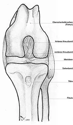
Skeleton
The vertebrae have muscles attached to the pedicles, the laminae, the spinous, transverse, and articular processes, the vertebral and intervertebral foramina, the atlas (C1), axis (C2), dens, and ventral lamina (C6).
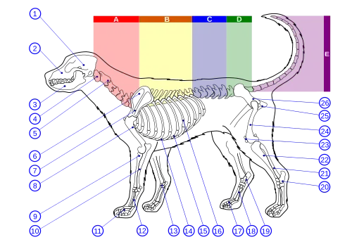
- Dog skeletal features
-
.jpg) Lateral view of a dog skeleton
Lateral view of a dog skeleton -
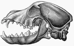 Lateral view of a dog skull, jaw opened
 -
_02.jpg) Lateral view of a dog skull, jaw closed
Lateral view of a dog skull, jaw closed -
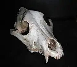 Frontal view of a dog skull
 -
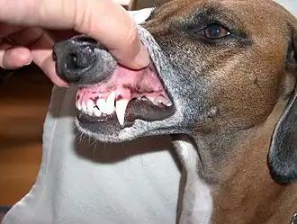 A dog's teeth
Skull
In 1986, a study of skull morphology found that the domestic dog is morphologically distinct from all other canids except the wolf-like canids. The difference in size and proportion between some breeds are as great as those between any wild genera, but all dogs are clearly members of the same species.[5] In 2010, a study of dog skull shape compared to extant carnivorans proposed that "The greatest shape distances between dog breeds clearly surpass the maximum divergence between species in the Carnivora. Moreover, domestic dogs occupy a range of novel shapes outside the domain of wild carnivorans."[6]
The domestic dog compared to the wolf shows the greatest variation in the size and shape of the skull (Evans 1979) that ranges from 7 to 28 cm in length (McGreevy 2004). Wolves are dolichocephalic (long-skulled) but not as extreme as some breeds of dogs, such as greyhounds and Russian wolfhounds (McGreevy 2004). Canine brachycephaly (short-skulledness) is found only in domestic dogs and is related to paedomorphosis (Goodwin 1997). Puppies are born with short snouts, with the longer skull of dolichocephalic dogs emerging in later development (Coppinger 1995). Other differences in head shape between brachycephalic and dolichocephalic dogs include changes in the craniofacial angle (angle between the basilar axis and hard palate) (Regodón 1993), morphology of the temporomandibular joint (Dickie 2001), and radiographic anatomy of the cribriform plate (Schwarz 2000).[7]
One study found that the relative reduction in dog skull length compared to its width (the cephalic index) was significantly correlated to both the position and the angle of the brain within the skull, regardless of the brain size or the body weight of the dog.[8]
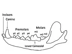
| Canid | Carnassial | Canine |
|---|---|---|
| Wolf | 131.6 | 127.3 |
| Dhole | 130.7 | 132.0 |
| African wild dog | 127.7 | 131.1 |
| Greenland Dog (domesticated) | 117.4 | 114.3 |
| Coyote | 107.2 | 98.9 |
| Side-striped jackal | 93.0 | 87.5 |
| Golden jackal | 89.6 | 87.7 |
| Black-backed jackal | 80.6 | 78.3 |
Respiratory system
The respiratory system is the set of organs responsible for the intake of oxygen and the expelling of carbon dioxide. As dogs have few sweat glands in their skin, the respiratory system also plays an important role in body thermoregulation.[10]
Dogs are mammals with two large lungs that are further divided into lobes. They have a spongy appearance due to the presence of a system of delicate branches of the bronchioles in each lung, ending in closed, thin-walled chambers (the points of gas exchange) called alveoli. The presence of a muscular structure, the diaphragm, exclusive to mammals, divides the peritoneal cavity from the pleural cavity, besides assisting the lungs during inhalation.
Inbreeding dogs can cause brachycephalic airway syndrome. The dog's face can have a shortened skull, facial and nasal bones, stenotic nares, a hypoplastic trachea, and everted laryngeal saccules.[11][12]
Digestive system
The organs that make up the canine digestive system are the same as those in most other mammals, including a mouth, esophagus, stomach, small and large intestines, rectum, anus, liver, and pancreas.[13]
-
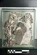
 Dog cecum
 -
 Dog digestive tract
 -
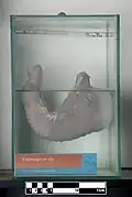 Dog stomach
 -
.jpg) Dog stomach (open, inner view)
Dog stomach (open, inner view) -
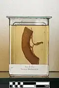
 Formalin fixed dog tongue
 -
 Segment of dog ileum
 -
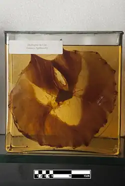
 Vascular structure of the dog liver
Reproductive system
Physical characteristics
Sixty percent of the dog's body mass falls on the front legs.[14]
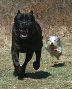
The dog has a cardiovascular system. The dog's muscles provide the dog with the ability to jump and leap. Their legs can propel them to leap forward rapidly to chase and overcome prey. They have small, tight feet and walk on their toes (thus having a digitigrade stance and locomotion). Their rear legs are fairly rigid and sturdy. The front legs are loose and flexible, with only muscle attaching them to the torso.
The dog's muzzle size will vary with the breed. Dogs with medium muzzles, such as the German Shepherd Dog, are called mesocephalic and dogs with a pushed in muzzle, such as the Pug, are called brachycephalic. Today's toy breeds have skeletons that mature in only a few months, while giant breeds, such as the Mastiffs, take 16 to 18 months for the skeleton to mature. Dwarfism has affected the proportions of some breeds' skeletons, as in the Basset Hound.
All living Canidae have a ligament connecting the spinous process of their first thoracic (or chest) vertebra to the back of the axis bone (second cervical or neck bone), which supports the weight of the head without active muscle exertion, thus saving energy.[15] This ligament is analogous in function (but different in exact structural detail) to the nuchal ligament found in ungulates.[15] This ligament allows dogs to carry their heads while running long distances, such as while following scent trails with their nose to the ground, without expending much energy.[15]
Dogs have disconnected shoulder bones (lacking the collar bone of the human skeleton) that allow a greater stride length for running and leaping. They walk on four toes, front and back, and have vestigial dewclaws on their front legs and on their rear legs. When a dog has extra dewclaws in addition to the usual one in the rear, the dog is said to be "double dewclawed."
Size
Dogs are highly variable in height and weight. The smallest known adult dog was a Yorkshire Terrier that stood only 6.3 cm (2.5 in) at the shoulder, 9.5 cm (3.7 in) in length along the head and body, and weighed only 113 grams (4.0 oz). The largest known adult dog was an English Mastiff, which weighed 155.6 kg (343 lb).[2] The tallest known adult dog is a Great Dane that stands 106.7 cm (42.0 in) at the shoulder.[3]
In 2007, a study identified a gene that was proposed to be responsible for dog size. The study found a regulatory sequence next to the gene Insulin-like growth factor 1 (IGF1), which, together with the gene and regulatory sequence, "is a major contributor to body size in all small dogs." Two variants of this gene were found in large dogs, making a more complex reason for the large breed size. The researchers concluded that this gene's instructions to make dogs small must be at least 12,000 years old and it is not found in wolves.[16] Another study has proposed that lap dogs (small dogs) are among the oldest existing dog types.[17]
Coat
Domestic dogs often display the remnants of countershading, a common natural camouflage pattern. The general theory of countershading is that an animal that is lit from above will appear lighter on its upper half and darker on its lower half, where it will usually be in its own shade.[18][19] This is a pattern that predators can learn to watch for. A counter-shaded animal will have dark coloring on its upper surfaces and light coloring below.[18] This reduces the general visibility of the animal. In this pattern, many breeds will have the occasional "blaze", stripe, or "star" of white fur on their chests or undersides.[19]
A study found that the genetic basis that explains coat colors in horse coats and cat coats did not apply to dog coats.[20] The project took samples from 38 different breeds to find the gene (a beta defensin gene) responsible for dog coat color. One version produces yellow dogs and a mutation produces black dogs. All dog coat colors are modifications of black or yellow.[21] For example, the white in white miniature schnauzers is a cream color, not albinism (a genotype of E/E' at MC1R).
Modern dog breeds exhibit a diverse array of fur coats, including dogs without fur, such as the Mexican Hairless Dog. Dog coats vary in texture, color, and markings, and a specialized vocabulary has evolved to describe each characteristic.[22]
Tail
There are many different shapes of dog tails: straight, straight up, sickle, curled and cork-screw. In some breeds, the tail is traditionally docked to avoid injuries (especially for hunting dogs).[23] It can happen that some puppies are born with a short tail or no tail in some breeds. The T-box gene mutation (C189G) is responsible for bobtail breeds having no tail to short tail.[24][25] Dogs have a violet gland or supracaudal gland on the dorsal (upper) surface of their tails.
Footpad
The dog's footpad is a fatty tissue locomotive-supporting organ, present at the bottom of the four legs, consisting of digital pads, a metacarpal pad, and a carpal pad, with dewclaw near the footpad.[26] When a dog's footpad is exposed to the cold, heat loss is prevented by an adaptation of the blood system that recirculates heat back into the body. It brings blood from the skin surface and retains warm blood on the pad surface.[27]
Senses
Vision
Like most mammals, dogs have only two types of cone photoreceptors, making them dichromats.[28][29][30][31] These cone cells are maximally sensitive between 429 nm and 555 nm. Behavioural studies have shown that the dog's visual world consists of yellows, blues and grays,[31] but they have difficulty differentiating between red and green, making their color vision equivalent to red–green color blindness in humans (deuteranopia). When a human perceives an object as "red," this object appears as "yellow" to the dog, and the human perception of "green" appears as "white," a shade of gray. This white region (the neutral point) occurs around 480 nm, the part of the spectrum that appears blue-green to humans. For dogs, wavelengths longer than the neutral point cannot be distinguished from each other, and all appear yellow.[31]
Dogs use color instead of brightness to differentiate between light or dark blue/yellow.[32][33][34] They are less sensitive to differences in gray shades than humans and can also detect brightness with about half the accuracy of humans.[35]: 140 The dog's visual system has evolved to aid in hunting.[28] Dogs have been shown to be able to discriminate between humans (e.g., identifying their human guardian) at a range of between 800 and 900 metres (2,600 and 3,000 ft); however, this range decreases to 500–600 metres (1,600–2,000 ft) if the object is stationary.[28] Dogs can detect a change in movement that exists in a single diopter of space within their eye. Humans, by comparison, require a change of between 10 and 20 diopters to detect movement.[36] A test has estimated poodles' visual acuity to have a Snellen rating of 20/75, a relatively low score compared to humans' vision.[28]
As crepuscular hunters, dogs often rely on their vision in low light situations: They have very large pupils, a high density of rods in the fovea, an increased flicker rate, and a tapetum lucidum.[28] The tapetum is a reflective surface behind the retina that reflects light to give the photoreceptors a second chance to catch the photons. There is also a relationship between body size and the overall diameter of the eye. A range of 9.5 and 11.6 mm can be found between various breeds of dogs. This 20% variance is associated with an adaptation toward superior night vision.[35]: 139
The eyes of different breeds of dogs have different shapes, dimensions, and retina configurations.[37] Many long-nosed breeds have a "visual streak"—a wide foveal region that runs across the width of the retina and gives them a very wide field of excellent vision. Some long-muzzled breeds, in particular, the sighthounds, have a field of vision up to 270° (compared to 180° for humans). Short-nosed breeds, on the other hand, have an "area centralis", a central patch with up to three times the density of nerve endings as the visual streak, giving them detailed sight much more like a human's. Some broad-headed breeds with short noses have a field of vision similar to that of humans.[29][30]
Most breeds have good vision, but some show a genetic predisposition for myopia—such as Rottweilers, with which one out of every two has been found to be myopic.[28] Dogs also have a greater divergence of the eye axis than humans, enabling them to rotate their pupils farther in any direction. The divergence of the eye axis of dogs ranges from 12–25°, depending on the breed.[36] Experimentation has found that dogs can distinguish between complex visual images such as those of a cube or a prism. Dogs also show attraction to static visual images such as the silhouette of a dog on a screen, their own reflections, or videos of dogs; however, their interest declines sharply once they are unable to make social contact with the image.[35]: 142
Hearing

The frequency range of dog hearing is between 16–40 Hz (compared to 20–70 Hz for humans) and up to 45–60 kHz (compared to 13–20 kHz for humans), which means that dogs can detect sounds beyond the upper limit of the human auditory spectrum.[30][38][39][40]
Dogs have ear mobility that allows them to rapidly pinpoint the exact location of a sound. Eighteen or more muscles can tilt, rotate, raise, or lower a dog's ear. A dog can identify a sound's location much faster than a human can, as well as hear sounds at four times the distance.[41] Dogs can lose their hearing from age or an ear infection.[42]
Smell
While the human brain is dominated by a large visual cortex, the dog brain is dominated by a large olfactory cortex.[28] Dogs have roughly forty times more smell-sensitive receptors than humans, ranging from about 125 million to nearly 300 million in some dog breeds, such as bloodhounds.[28]
Taste
Dogs have around 1,700 taste buds compared to humans, with around 9,000. The sweet taste buds in dogs respond to furaneol. It appears that dogs do like this flavor, and it probably evolved because, in a natural environment, dogs frequently supplement their diet of small animals with whatever fruits are available. Because of dogs' dislike of bitter tastes, various sprays, and gels have been designed to keep dogs from chewing on furniture or other objects. Dogs also have taste buds that are tuned for water, which is something they share with other carnivores but is not found in humans. This taste sense is found at the tip of the dog's tongue, which is the part of the tongue that they curl to lap water. This area responds to water at all times, but when the dog has eaten salty or sugary foods, the sensitivity to the taste of water increases. It is proposed that this ability to taste water evolved as a way for the body to keep internal fluids in balance after the animal has eaten things that will either result in more urine being passed or will require more water to adequately process. It appears that when these special water taste buds are active, dogs seem to get an extra pleasure out of drinking water, and will drink copious amounts of it.[43]
Touch
Dogs have specialized whiskers known as vibrissae, sensing organs present above the dog's eyes, below their jaw, and on their muzzle. Vibrissae are more rigid, embedded much more deeply in the skin than other hairs, and have a greater number of receptor cells at their base. They can detect air currents, subtle vibrations, and objects in the dark. They provide an early warning system for objects that might strike the face or eyes, and probably help direct food and objects towards the mouth.[44]
Magnetic sensitivity
A study found that dogs may prefer, when they are off the leash and the Earth's magnetic field is calm, to urinate and defecate with their bodies aligned on a north-south axis. Dogs are sensitive to changes in the Earth's magnetic field polarity.[45] No significant differences between males and females in angular preferences were found. Some studies have detected cryptochrome 1 in some dogs' photoreceptors' blue-sensitive cones.[46][47]
Temperature regulation
Primarily, dogs regulate their body temperature through panting[48] and sweating via their paws. Panting moves cooling air over the moist surfaces of the tongue and lungs, transferring heat to the atmosphere.
Dogs and other canids also possess a set of nasal turbinates, an elaborate set of bones and associated soft-tissue structures (including arteries and veins) in the nasal cavities. These turbinates allow for heat exchange between small arteries and veins on their maxilloturbinate surfaces (the surfaces of turbinates positioned on maxilla bone) in a counter-current heat-exchange system. Compared to the ambush predation of cats, dogs are capable of prolonged chases due to these turbinates (cats possess a much smaller and less-developed set of nasal turbinates).[49]: 88 This same turbinate structure helps conserve water in arid environments. The water conservation and thermoregulatory capabilities of these turbinates in dogs may have allowed dogs (including both domestic dogs and their wild prehistoric ancestors) to survive in the Arctic environment and other cold areas of northern Eurasia and North America, which are dry and cold.[49]: 87
References
- ^ Scientists fetch useful information from dog genome publications, Cold Spring Harbor Laboratory, 7 December 2005; published online in Bio-Medicine Archived 19 November 2020 at the Wayback Machine quote: "Phenotypic variation among dog breeds, whether it be in size, shape, or behavior, is greater than for any other animal"
- ^ a b Donald McFarlan (1 December 1988). Guinness Book of World Records, 1989. Sterling. p. 47. ISBN 978-0-8069-0276-0. Retrieved 14 November 2012.
- ^ a b "Guinness World Records – Tallest Dog Living". Guinness World Records. 31 August 2004. Archived from the original on 11 July 2011. Retrieved 7 January 2009.
- ^ Evans, Howard E.; de Lahunta, Alexander (2017). Guide to the Dissection of the Dog (8th ed.). St. Louis, Missouri: Elsevier. ISBN 978-0-323-39165-8. OCLC 923139309.
- ^ Wayne, Robert K. (1986). "Cranial Morphology of Domestic and Wild Canids: The Influence of Development on Morphological Change". Evolution. 40 (2): 243–261. doi:10.2307/2408805. JSTOR 2408805. PMID 28556057.
- ^ Drake, Abby Grace; Klingenberg, Christian Peter (2010). "Large-Scale Diversification of Skull Shape in Domestic Dogs: Disparity and Modularity". The American Naturalist. 175 (3): 289–301. Bibcode:2010ANat..175..289D. doi:10.1086/650372. PMID 20095825. S2CID 26967649.
- ^ Roberts, Taryn; McGreevy, Paul; Valenzuela, Michael (2010). "Human Induced Rotation and Reorganization of the Brain of Domestic Dogs". PLOS ONE. 5 (7): e11946. Bibcode:2010PLoSO...511946R. doi:10.1371/journal.pone.0011946. PMC 2909913. PMID 20668685. All cited in Roberts.
- ^ Roberts, Taryn; McGreevy, Paul; Valenzuela, Michael (2010). "Human Induced Rotation and Reorganization of the Brain of Domestic Dogs". PLOS ONE. 5 (7): e11946. Bibcode:2010PLoSO...511946R. doi:10.1371/journal.pone.0011946. PMC 2909913. PMID 20668685.
- ^ Christiansen, Per; Wroe, Stephen (2007). "Bite Forces and Evolutionary Adaptations to Feeding Ecology in Carnivores". Ecology. 88 (2): 347–358. doi:10.1890/0012-9658(2007)88[347:bfaeat]2.0.co;2. PMID 17479753.
- ^ Washington State University. "Respiratory System of the Dog". Retrieved 1 June 2017.Archived 2016-11-08 at the Wayback Machine
- ^ "Brachycephalic Airway Syndrome in Dogs". www.petmd.com. Retrieved 28 March 2024.
- ^ Ravn-Mølby, Eva-Marie; Sindahl, Line; Nielsen, Søren Saxmose; Bruun, Camilla S.; Sandøe, Peter; Fredholm, Merete (16 December 2019). "Breeding French bulldogs so that they breathe well—A long way to go". PLOS ONE. 14 (12): e0226280. Bibcode:2019PLoSO..1426280R. doi:10.1371/journal.pone.0226280. ISSN 1932-6203. PMC 6913956. PMID 31841527.
- ^ Washington State University. "Digestive System of the Dog". Retrieved 31 May 2017.
- ^ Fish, Frank E.; Sheehan, Maura J.; Adams, Danielle S.; Tennett, Kelsey A.; Gough, William T. (2021). "A 60:40 split: Differential mass support in dogs". The Anatomical Record. 304 (1): 78–89. doi:10.1002/ar.24407. PMID 32363786. S2CID 218491599.
- ^ a b c Wang, Xiaoming and Tedford, Richard H. Dogs: Their Fossil Relatives and Evolutionary History. New York: Columbia University Press, 2008. pp.97-8
- ^ Sutter NB, Bustamante CD, Chase K, et al. (April 2007). "A single IGF1 allele is a major determinant of small size in dogs". Science. 316 (5821): 112–5. Bibcode:2007Sci...316..112S. doi:10.1126/science.1137045. PMC 2789551. PMID 17412960.
- ^ Ostrander EA (September–October 2007). "Genetics and the Shape of Dogs; Studying the new sequence of the canine genome shows how tiny genetic changes can create enormous variation within a single species". Am. Sci. Archived from the original on 27 April 2012. Retrieved 6 November 2008.
- ^ a b Klappenbach, Laura (2008). "What is Counter Shading?". About.com. Archived from the original on 27 September 2011. Retrieved 22 October 2008.
- ^ a b Cunliffe, Juliette (2004). "Coat Types, Colours and Markings". The Encyclopedia of Dog Breeds. Paragon Publishing. pp. 20–3. ISBN 0-7525-6561-3.
- ^ Candille SI, Kaelin CB, Cattanach BM, et al. (November 2007). "A -defensin mutation causes black coat color in domestic dogs". Science. 318 (5855): 1418–23. doi:10.1126/science.1147880. PMC 2906624. PMID 17947548.
- ^ Stanford University Medical Center, Greg Barsh et al. (2007, 31 October). Genetics Of Coat Color In Dogs May Help Explain Human Stress And Weight. ScienceDaily. Retrieved 29 September 2008
- ^ "Genetics of Coat Color and Type in Dogs". Sheila M. Schmutz, Ph.D., Professor, University of Saskatchewan. 25 October 2008. Archived from the original on 14 February 2020. Retrieved 5 November 2008.
- ^ "The Case for Tail Docking". cdb.org. Archived from the original on 14 April 2009. Retrieved 22 October 2008.
- ^ Hytönen, Marjo K.; Grall, Anaïs; Hédan, Benoît; Dréano, Stéphane; Seguin, Samuel J.; Delattre, Delphine; Thomas, Anne; Galibert, Francis; Paulin, Lars; Lohi, Hannes; Sainio, Kirsi; André, Catherine (2009). "Ancestral T-box mutation is present in many, but not all, short-tailed dog breeds". The Journal of Heredity. 100 (2): 236–240. doi:10.1093/jhered/esn085. ISSN 1465-7333. PMID 18854372.
- ^ "Dog Breeds Born Without Tails". 14 August 2019.
- ^ Johansen, Kyle (October 2024). "4 Things You Should Know About Your Dog's Paws". How I Met My Dog.
- ^ Ninomiya, Hiroyoshi; Akiyama, Emi; Simazaki, Kanae; Oguri, Atsuko; Jitsumoto, Momoko; Fukuyama, Takaaki (2011). "Functional anatomy of the footpad vasculature of dogs: Scanning electron microscopy of vascular corrosion casts". Veterinary Dermatology. 22 (6): 475–81. doi:10.1111/j.1365-3164.2011.00976.x. PMID 21438930.
- ^ a b c d e f g h Coren, Stanley (2004). How Dogs Think. First Free Press, Simon & Schuster. ISBN 0-7432-2232-6.
- ^ a b A&E Television Networks (1998). Big Dogs, Little Dogs: The companion volume to the A&E special presentation. A Lookout Book. GT Publishing. ISBN 1-57719-353-9.
- ^ a b c Alderton, David (1984). The Dog. Chartwell Books. ISBN 0-89009-786-0.
- ^ a b c Jennifer Davis (1998). "Dr. P's Dog Training: Vision in Dogs & People". Archived from the original on 9 February 2015. Retrieved 20 February 2015.
- ^ Anna A. Kasparson; Jason Badridze; Vadim V. Maximov (July 2013). "Colour cues proved to be more informative for dogs than brightness". Proceedings of the Royal Society B: Biological Sciences. 280 (1766): 20131356. doi:10.1098/rspb.2013.1356. PMC 3730601. PMID 23864600.
- ^ Jay Neitz; Timothy Geist; Gerald H. Jacobs (1989). "Color Vision in the Dog" (PDF). Visual Neuroscience. 3 (2): 119–125. doi:10.1017/s0952523800004430. PMID 2487095. S2CID 23509491. Archived from the original (PDF) on 13 April 2015. Retrieved 23 June 2015.
- ^ Jay Neitz; Joseph Carroll; Maureen Neitz (January 2001). "Color Vision — Almost Reason Enough for Having Eyes" (PDF). Optics & Photonics News. 12 (1): 26–33. Bibcode:2001OptPN..12...26N. doi:10.1364/OPN.12.1.000026. Archived from the original (PDF) on 4 March 2016. Retrieved 23 June 2015.
- ^ a b c Miklósi, Adám (2009). Dog Behaviour, Evolution, and Cognition. Oxford University Press. doi:10.1093/acprof:oso/9780199295852.001.0001. ISBN 978-0-19-929585-2.
- ^ a b Mech, David. Wolves, Behavior, Ecology, and Conservation. The University of Chicago Press, 2006, p. 98.
- ^ Jonica Newby; Caroline Penry-Davey (25 September 2003). "Catalyst: Dogs' Eyes". Australian Broadcasting Corporation. Retrieved 26 November 2006.
- ^ Elert, Glenn; Timothy Condon (2003). "Frequency Range of Dog Hearing". The Physics Factbook. Retrieved 22 October 2008.
- ^ "How well do dogs and other animals hear". Archived from the original on 28 August 2011. Retrieved 7 January 2008.
- ^ "How well do dogs and other animals hear".
- ^ "Dog Sense of Hearing". seefido.com. Archived from the original on 1 May 2009. Retrieved 22 October 2008.
- ^ Gibeault, Stephanie (22 February 2024). "Dogs Don't Have a Sixth Sense, They Just Have Incredible Hearing". American Kennel Club. Retrieved 28 March 2024.
- ^ Coren, Stanley
- ^ , Santos, A "Puppy and Dog Care: An Essential Puppy Training Guide", 2015 Amazon Digital Services, Inc. [1] Archived 24 June 2015 at the Wayback Machine
- ^ Hart, V.; Nováková, P.; Malkemper, E.; Begall, S.; Hanzal, V. R.; Ježek, M.; Kušta, T. Š.; Němcová, V.; Adámková, J.; Benediktová, K. I.; Červený, J.; Burda, H. (2013). "Dogs are sensitive to small variations of the Earth's magnetic field". Frontiers in Zoology. 10 (1): 80. doi:10.1186/1742-9994-10-80. PMC 3882779. PMID 24370002.
- ^ Magnetoreception molecule found in the eyes of dogs and primates Archived 23 December 2016 at the Wayback Machine MPI Brain Research, 22 February 2016
- ^ Nießner, Christine; Denzau, Susanne; Malkemper, Erich Pascal; Gross, Julia Christina; Burda, Hynek; Winklhofer, Michael; Peichl, Leo (2016). "Cryptochrome 1 in Retinal Cone Photoreceptors Suggests a Novel Functional Role in Mammals". Scientific Reports. 6 (1): 21848. Bibcode:2016NatSR...621848N. doi:10.1038/srep21848. PMC 4761878. PMID 26898837.
- ^ "How do Dogs Sweat?". Archived from the original on 14 December 2013. Retrieved 25 June 2010.
- ^ a b Wang, Xiaoming (2008) Dogs: Their Fossil Relatives and Evolutionary History Columbia University Press. ISBN 978-0-231-50943-5
Further reading
- Klaus-Dieter Budras (7 December 2010). Anatomy of the Dog: With Aaron Horowitz and Rolf Berg. Schlütersche Verlagsgesellschaft mbH & Company KG. ISBN 978-3-89993-099-3.
- Horowitz, Alexandra (2009). Inside of a Dog: What Dogs See, Smell, and Know. New York: Charles Scribner's Sons. ISBN 978-1-4165-8340-0. OCLC 973655798. Inside of a Dog: What Dogs See, Smell, and Know at Google Books.
- Howard E. Evans; Alexander de Lahunta (7 August 2013). Miller's Anatomy of the Dog - E-Book. Elsevier Health Sciences. ISBN 978-0-323-26623-9.
External links
Anatomy and morphology | ||
|---|---|---|
| Fields | 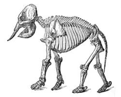 | |
| Bacteria and fungi | ||
| Protists |
| |
| Plants | ||
| Invertebrates | ||
| Mammals | ||
| Other vertebrates | ||
| Glossaries | ||
| Related topics | ||
| ||
| Types | |
|---|---|
| Breeds | |
| Roles | |
| Behavior | |
| Human–dog interaction | |
| Health | |
| Training | |
| Related | |
| |
.jpg)